Three-dimensional segmentation and reconstruction of medical images is currently
one of the most researched areas of image processing and analysis. There is a growing
need for automated tools that can reconstruct structures in three dimensions from
complex medical images, to make it easier to diagnose and study disease.
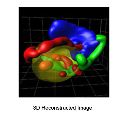
This project aims at studying the hemoglobin and nutrient uptake by the malarial parasite,
which is one of the most investigated topics in Plasmodium biology and is one of the greatest
potential drug targets. The images being used for this project are serial Transmission
Electron Micrographs (TEM) of the malarial parasite inside the human red blood cell. Each
dataset contains approximately 40 slices (100 nm thick) of the parasite. The goal of this
project is to reconstruct in 3-D the malarial parasite and hence determine the size of its
food vacuole and the volume of hemoglobin taken up by it. Analysis of reconstructed 3-D images
can differentiate between decreased uptake due to slower growth and decreased uptake due to
altered kinetics of a process.
Shown below are the raw image, the colored segmented image, and the 3-D reconstructed image.

This work is a collaborative effort with Prof. David A. Elliott in the
Department of Cell Biology and Anatomy, College of Medicine,
University of Arizona.
Publications:
-
Sunil Seepuri, Jeffrey J. Rodriguez and David A. Elliot, "Automated 3-D
Segmentation of Internal Haemoglobin in TEM Images" 2008 IEEE Southwest Symp. on Image Analysis and
Interpretation, March 24-26, 2008, Santa fe, NM, pp. 117-120. [ PDF ]