Accurate segmentation of breast cancer tissues stained with the help of a coloring agent is
important to aid the oncologist in researching the effects of the tumor on the blood vasculature
surrounding it. These are high-resolution color images of histological slides
The objective of this project is to develop an automated system to segment these regions on
interest accurately. Since the coloring agent is retained by the regions of interest, there is
a corresponding variation in color between these regions and the background. Principal component
analysis using the Karhunen-Loeve Transform is performed first in order to find features to
classify the regionsof interest from background. The luminance information is contained in
the first principal component and the chrominance information in the second and third principal
component. We consider the second principal component as the third principal component is
weak in energy.
The segmentation is performed by a seeded region growing scheme. The seed pixels within each
region of interest is are generated by thresholding the second principal component image. This
produces a lot of seed pixels which are pruned using a seed pruning stage. Once the final number
of seed pixels are found out, each if these are considered as starting point and multiple regions
are grown within a single image using the intensity of the second principal component as the
homogeneity criterion for growing the region.
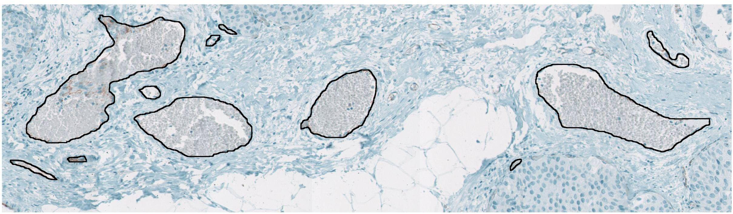
Figure 1: Input image showing manually outlined ROIs.
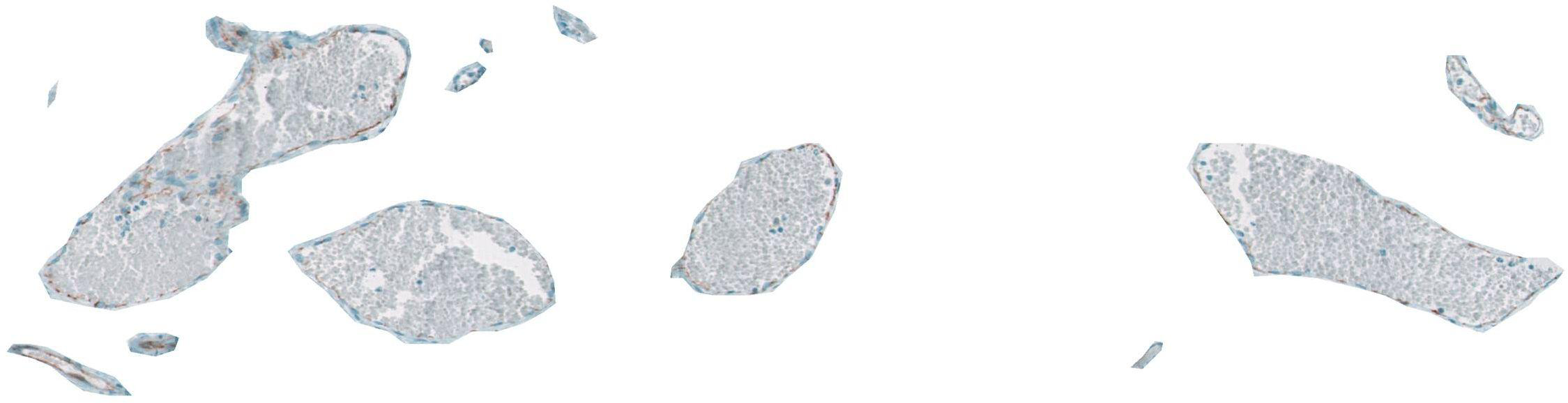
Figure 2: Automated segmentation using our approach.
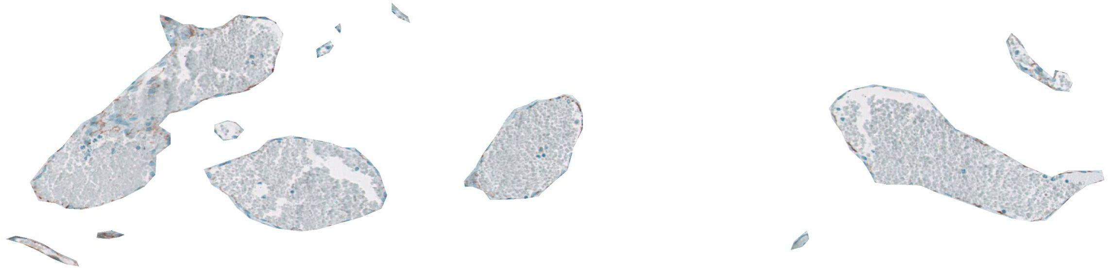
Figure 3: Manual segmentation.
This work is a collaborative effort with Prof. Robert J. Gillies (
Dept. of Biochemistry and Arizona Cancer Center
)
Publications:
-
Rohit C. Philip, Jeffrey J. Rodriguez, and Robert J. Gillies,
"Seed pruning using a multi-resolution approach for automated
segmentation of breast cancer tissue," 2008 IEEE Intl. Conf.
on Image Processing, San Diego, CA, Oct. 12-15, 2008, pp. 1436-9. [ PDF ]