Recent technological developments in the field of microscopy have brodened
the scope of digital image analysis in biological applications. Molecular
biologists are now able to image sub-nuclear structures in 3-D within a
resolution of about a few hundred nanometers. With the ability to probe
into the structure of the cells, researches hope to find answers to some of
the most intriguing questions in the field of molecular biology.
This project aims at quantifying the respose of HeLa
cells to varying doses of UV radiation. The response of a particular DNA
replication protein, RPA, is studied in detail. The goal is to find the
number, size and intensity of RPA molecules that congregate at sites of
damage in the DNA. We also wish to study the co-localization of the
different subunits of RPA at damage sites.
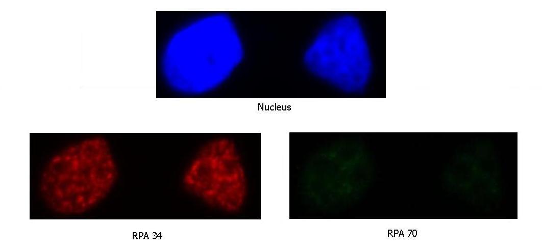
Images obtained before UV radiation (center slice from a 3-D stack):
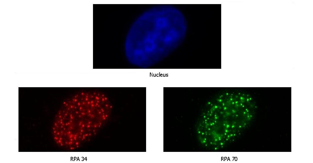
Images obtained after UV radiation (center slice from a 3-D stack):
This work is a collaborative effort with Prof. Kathleen Dixon in the
Dept. of Molecular and Cellular Biology, College of Science,
University of Arizona.
Publications:
-
Narasiman Rajagopalan, Jeffrey J. Rodriguez, and Kathleen Dixon, "An
integrated technique for volume estimation of spots in 3-D human cell cultures: Watersnakes
," 2008 IEEE Intl. Conf. on Image Processing, San Diego, CA, Oct. 12-15, 2008,
pp. 3004-3007. [ PDF ]