Digital imaging techniques have been considered for use in automatic karyotyping
of human chromosomes, but this is a new area where much work is still needed.
In this project, specific image analysis techniques were developed and
tested for automatic segmentation of a metaphase image of chromosomes.
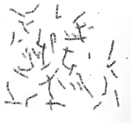
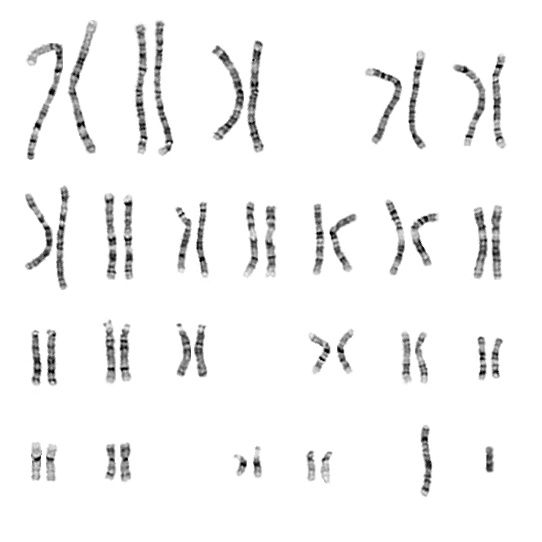
Typical chromosome image and the corresponding karyotype image:
A human chromosome is a highly structured, dynamic complex made of DNA and
protein. A normal diploid cell is supposed to have exactly 22 pairs of
chromosomes, 22 pairs of autosomes, and 2 sex chromosomes. If there is even
a small deviation from these numbers, then physical abnormalities can result.
About 6% of congenital abnormalities are due to chromosome anomalies.
Digital imaging has been used to capture metaphasic chromosomes
that have been marked with a specific dye. The goal in this project was to develop automatic
digital image analysis techniques to aid in karyotyping of the chromosomes.
Karyotyping is the process of grouping the chromosomes into pairs, based on
their identifying characteristics, and then presenting the pairs in an image
display (see the example below). Medical specialists use this display of
karyotyped chromosomes to diagnose abnormalities in patients. Scientists use
the karyotype for research in heredity and related topics. The study of
chromosomes and their abnormalities is referred to as cytogenetics. The major
difficulty in cytogenetics is the time-consuming nature of the manual
karyotyping process. If this manual stage can be automated, then diagnosis
and cytogenetic research would be greatly facilitated.
This work was a collaborative effort with Dr. Christopher M. Cunniff
in the Dept. of Pediatrics at University of Arizona.
Publications:
-
Victor Gajendran and Jeffrey J. Rodriguez,
"Chromosome Counting via Digital Image Analysis," in
Proc. 2004 IEEE Intl. Conf. on Image Processing,
Oct. 24-27, 2004, Singapore, vol. 5, pp. 2929-2932. [ PDF ]