Magnetic resonance imaging, computed tomography, mammography,
ultrasound and other digital imaging techniques are invaluable tools for
medical applications today. These techniques have been used to map the
anatomy of individuals, thus increasing our knowledge of disease, and hence
are used for diagnosis and treatment. Image segmentation algorithms are
used for the automatic delineation of anatomical structures or other
regions of interest in a digital image. There is no single acceptable
segmentation algorithm for all applications. General methods exist which
can be applied to a variety of data. However, using prior knowledge about
the data for which the application is intended can enhance performance.
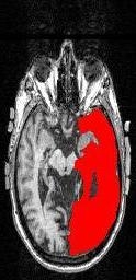
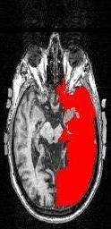
The focus of this work was to develop a scheme to automatically detect lesions
in T1-weighted MR images of patients with a history of stroke. Manual
segmentation is the most accurate technique to map the lesions in the brain
and is considered to be the gold standard. Thus, an automated segmentation
algorithm was developed in this project. This approach leads to
huge savings in the time required to map the lesions compared to the manual
tracing method. The performance was compared with results obtained by
manual segmentation and we observed improvements
in accuracy and speed compared with the most current region-growing algorithm.
Here is an example of (a) an image containing a lesion, along with
(b) the results of manual segmentation and (c) automated segmentation:
This work was a collaborative effort with Prof. Pélagie Beeson
in the Dept. of Speech & Hearing Sciences.