|
Past Research |
Texture Analysis of Optical-Coherence Tomographic Images
Student: Kirk W. Gossage
| |
Optical coherence tomography (OCT) acquires cross-sectional images of
tissue by measuring back-reflected light. Images from in vivo OCT systems
typically have a resolution of 10-15mm, and are thus best suited for
visualizing features in the tens to hundreds of microns size range such as
tissue layers or glands. Many normal and abnormal tissues lack features in
this size range so it may appear that OCT is unsuitable for identification
of these tissues. However, examination of feature-poor OCT images reveals
that they frequently display a characteristic repetitive structure due to
speckle.
The purpose of this study was to evaluate the application of statistical and
spectral texture analysis techniques for differentiating tissue types based
on the structural and speckle content in OCT images.
Experiments yielded promising classification rates, demonstrating
that texture analysis of OCT images may be capable of
differentiating tissue types in the absence of visibly identifiable
structures.
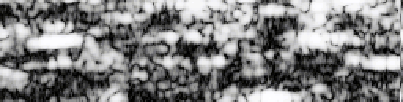
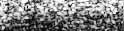
Here are some example OCT images of mouse fat and lung, respectively:
Principal dissertation advisor:
Prof. Jennifer K. Barton, Biomedical Engineering.
Publications:
-
Kirk W. Gossage, Cynthia M. Smith, Elizabeth M. Kanter,
Lida P. Hariri, Alice L. Stone, Jeffrey J. Rodriguez,
Stuart K. Williams, and Jennifer K. Barton,
"Texture Analysis of Speckle in Optical
Coherence Tomography Images of Tissue Phantoms,"
Physics in Medicine and Biology, vol. 51, no. 6,
March 21, 2006, pp. 1563-1575. [ PDF ]
-
Kirk W. Gossage, Cynthia M. Smith, Elizabeth M. Kanter, Lida P. Hariri,
Alice L. Stone, Jeffrey J. Rodriguez, Stuart K. Williams, and
Jennifer K. Barton, "Texture Analysis of Speckle in Optical Coherence
Tomography Images of Tissue Phantoms," in
Advanced Biomedical and Clinical Diagnostic Systems II,
Gerald E. Cohn et al., Eds., Proc. SPIE, vol. 5318, 2004, pp.
140-150.Presented at Photonics West, San Jose, CA, Jan. 25-26, 2004. [ PDF ]
-
Kirk W. Gossage, Tomasz S. Tkaczyk, Jeffrey J. Rodriguez, and
Jennifer K. Barton, "Texture Analysis of Optical Coherence
Tomography Images: Feasibility for Tissue Classification,"
Journal of Biomedical Optics, vol. 8, no. 3, July 2003, pp.
570-575. [ PDF ]
-
Kirk W. Gossage, Tomasz S. Tkaczyk, Jeffrey J. Rodriguez, and
Jennifer K. Barton, "Texture Analysis for Tissue Classification of
Optical Coherence Tomography Images," in Advanced Biomedical
and Clinical Diagnostic Systems, Tuan Vo-Dinh et al., Eds. Proc.
SPIE, vol. 4958, 2003, pp. 109-17. Presented at the Biomedical Optics
Symp. (BiOS 2003) at Photonics West 2003, San Jose, CA, Jan. 26-28,
2003. [ PDF ]
|
|