
Department of Electrical & Computer Engineering
Signal and Image Laboratory (SaIL)
The University of Arizona®
|
|
|
|
Past Research |
Analysis of Image Quality of Medical Imaging Displays
Student: Amarpreet S. Chawla
| |
In most radiological imaging workstations today, soft-copy diagnosis is
done on a monochrome cathode ray tube (CRT) display or on a liquid
crystal display (LCD). The relevance of quality control of these imaging systems
has been well recognized and several methods have been used for performing
quality evaluation. These methods can be broadly classified into psychophysical
and analytical studies.
In this project, we developed more comprehensive comprehensive methods to evaluate the
transfer function and to refine the existing techniques. Work was also done on quantifying
the noise (including spatial and temporal components) on these displays and finding a
relationship with results from the psychophysical studies. The ultimate aim is to
devise a display system on which the image perception is not distorted due
to the inherent properties of the display and effects from the human eye.
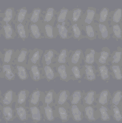
The following image shows LCD pixels magnified, in which
the three components (subpixels) of each
pixel and the spatial noise are clearly visible.
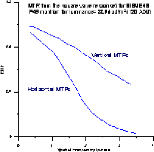
Also shown is a typical transfer function (horizontal and vertical)
for an LCD display.
Publications:
-
Amarpreet S. Chawla, Hans Roehrig, Jeffrey J. Rodriguez, and
Jiahua Fan, "Determining the MTF of Medical Imaging Displays Using
Edge Techniques," Journal of Digital Imaging, vol. 18, no.
4, Dec. 2005, pp. 296-310. [ PDF ]
|
|
1230 E. Speedway Blvd., P.O. Box 210104, Tucson, AZ 85721-0104
|
©2014 All Rights Reserved. |
| Contact webmaster
|
|
|
|
|